Laboratory of Microscopy
An expertise at the micro/nano scale
- To visualise and examine the lignocellulosic materials
- To identify the fibres and the minerals used in biosourced materials
- To understand the impact of production processes on the fibres and their derivatives
- To analyse the production defaults
- To produce some microphotographs to illustrate the scientific results
General informations
The laboratory of microscopy is a support for the industrialists and the research CTP teams to understand the processes and the phenomena observed as well as during research or during industrial production. The use of microscopy allows understanding the mechanisms, identifying and localizing the fibrous elements and the minerals and to analyse the production defaults.The samples to analyse are numerous:
- Organic, mineral or microbiological deposits,
- Monolayer or multilayers cellulosic, mineral and organic supports
- Papers, boards, moulded objects, wood, annual plants and lignocellulosic or synthetic fibres.
- Nanometric, inorganics or lignocellulosic objects, of which microfibrillated cellulose (MFC).
Available Equipments
Binocular Magnifier
SteREO Discovery V8, Zeiss (2007)
- Wild-field imaging
- Zoom x1 to x8
- Camera
Light microscope
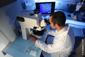
Axio Imager Z2, Zeiss (2013)
- Transmission lenses from x5 to x100
- Reflection lenses from x10 to x50
- Motorised turntable in XYZ
- Fluorescence light
- High resolution camera (pixel size 3.45 μm x 3.45 μm)
Digital Microscope
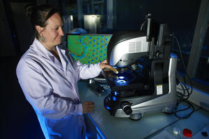
VHX-7000, Keyence (2023)
- Objectives from X20 to x2500
- Motorised stage in XYZ Large depth of field : 3D images
- Various lighting modes (annular, transmission, coaxial)
- 3D enhancement
- Self-assembly of images, video
- Profile, height and volume data
- Surface roughness
Scanning Electron Microscope - MEB-FEG
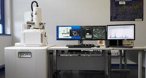
JSM-IT500HR LV, JEOL (2018-CMTC)
-Field effect gun (FEG)
-High vacuum mode (10-4PA) or low vacuum mode (10-150 PA)
- Acceleration tension: 0.5 to 30 KV
- Detector of secondary electrons
- Detector of retro-diffused electrons
- SDD detector for X microanalysis with energy dispersion (EDS)
- Topographic mode/composition/cartography
 |
 |
 |
||
| Digital Microscope, Keyence VHX7000 (2023) | Light microscope-AXIO Imager Z2, Zeiss (2013) | MEB-JSM-IT500HR, JEOL (2018, CMTC) |